Environmental Toxicity: An Alternative Way of Assessing Heavy Metals |
by Dr. Thomas Nissen |
Numerous scientists world wide are supporting the view today that all life processes are being determined by subtle electromagnetic and photon phenomena [see Prof. Dr. A. Popp,, Dr. Voll (EAP), Dr. Dr. Schimmel (Vega System) and many more). All electrically active metals (ions) and particularly heavy metals, can disturb the harmony of the electromagnetic and photon energies in our body, causing disharmony and disease. They also can increase the production of free radicals million-fold. It has been stated that 90 % of all chronic and serious illnesses could be prevented if we were able to eliminate the 600 most dangerous environmental toxins (Dr. J. Higgensen, Head of Cancer Research, WHO, Geneva, Switzerland). Every health practitioner is fully aware of the devastating influence heavy metals and/or ionic metals can have on our mental, emotional and physical health and well being. Until recently, most health care professionals and researchers assumed that heavy metals had to be taken into account only when a patient showed definite symptoms of 'poisoning'. We realize now that our health and well-being is affected by much lower levels of heavy metals than previously assumed. Health authorities constantly correct 'permissible' maximum levels downwards. It is becoming more difficult to accurately determine the appropriate drug profile in a given case, because the respective simile of symptoms has undergone a shift due to the presence of heavy metal ions. In fact, this phenomenon may be observed for the majority of the classic Hahnemann remedy profiles and it is fair to say that at the present time the effectiveness of any antioxidant therapy is significantly compromised by the presence of heavy metal ions. In cases of acute heavy metal poisoning (commonly the result of accidents or extreme workplace related contamination), clinical toxicology is generally able to provide an effective quick response with the DMPS procedure administered as mobilization test and antidote. However, hardly any appropriate treatment or diagnostic procedure is available for cases of long-term heavy metal contamination. No satisfactory method exists for the early recognition of heavy metal contamination. |
| Two Types of Metals |
The methods used to detect heavy metal contamination are cumbersome and costly and in some instances can’t differentiate between organically bound and free metal atoms (e.g. Cu, Zn in spectrometric analyses). Recent research has shown that it is essentially electrically active heavy metal atoms not bound with organic complexes that actively destroys molecular compounds and thereby cause the formation of free radicals. Unfortunately, traditional methods like hair or blood analyses are not able to uncover these connections for the simple reason that the organic sample is destroyed in the course of the analysis. Such procedures are therefore unable to differentiate between metal atoms bound with organic complexes and unbound and therefore electro-magnetically active ions, a difference that is crucial in the assessment of the overall situation. |
| A New Way to Assess Heavy Metals |
In 1925 Helmut Fischer of the Siemens Concern in Berlin succeeded in detecting heavy metal ions by means of a dithizone process. As a reagent, dithizone is able to indicate the presence of heavy metal ions in qualitative and in quantitative terms. In binding with them, colored complexes are formed in the interior of the molecule which are soluble in non polar organic solvents. The coloration of these solutions is very intensive, its particular coloration determined by the atomic radius of the respective metal present in the complex. The dithizon heavy metal reagent allows the detection of free heavy metal ions in bodily liquids like urine and saliva . By administering the test reagent as an exploratory measure, contaminations from amalgam fillings or from the environment (cadmium, lead, zinc, copper, manganese, nickel and cobalt - pointing to infections, organ or system disorders) can be identified on the spot, the potential health problem, as well as the need for detoxification before any specific therapy is administered. The dithizone reagent can also be used to determine the environmental sources of the contamination in aqueous solutions such as tap water and since all heavy metal ions are water soluble, solids like food items, porcelain dishes, dust samples from carpets, wall paints and wall paper etc. can be tested for heavy metals by soaking them in distilled water beforehand. |
| Replacement Reaction or How to Assess Mercury Toxicity |
The sheep study done at the University of Calgary in Canada(sheep had amalgam fillings placed in their mouths) clearly shows that very little mercury is found in the urine and in the blood, but highest amount are shown in the kidneys. Since this is the case how to assess mercury toxicity via the urine? In the human system, the bivalent metals are engaged in a continuous fight against one another, e.g. copper against zinc, magnesium against calcium, which results in the replacement of the "lighter" element by the "heavier" one in terms of their atomic masses. Replacement reactions, also called fight for the site, occur when heavy metals grab the biological spaces that should be filled by necessary minerals. Just as carbon monoxide replaces essential oxygen, other elements and compounds cause their toxic effect by replacing chemicals essential to the body functions. Within a group, for example group 2 in the periodic table of elements( 2 refers to the number of extra electron) there is zinc (Zn), cadmium (Cd), and mercury (Hg), in order of increasing atomic weight. (65, 112, and 200 respectively). Since cadmium and mercury, in their more soluble ionized or salt forms, will attempt to participate in the same biochemical reactions as zinc, their presence will prevent the zinc reacting and performing its functions in the body. This is like a 65 pound person (zinc) competing unsuccessfully with 112 pound (cadmium) and 200 pound (mercury) people in a game of musical chairs. Other symptoms caused by mineral deficiency and displacement by a heavy metal. (Hg, Cd, Pb, ) include: |
|
| Causing a Toxic Accumulation of Essential Minerals |
By taking the biological spaces of the essential minerals, heavy metals, in particular mercury, create simultaneously a toxic accumulation of essential minerals. The body receives everyday essential minerals through the food, unable to be absorbed leading to an accumulation and overburden of these minerals. |
| Case Study |
Here are the results of one case that shows the importance of heavy metal assessment: 14 year old male, with very advanced 3rd degree scoliosis (adequate amounts of copper are required for the normal production of elastin and collagen, which are the primary components of ligaments and the spinal discs), shy and timid. The parents (father is a lawyer) are healthy and live in a good environment. The urine revealed very high amounts of free copper and zinc ions (unbound). |
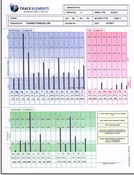
Highly Recommended: One thing we recommend for anyone who is experiencing autoimmune symptoms is to have a hair tissue mineral analysis (HTMA) to test for heavy metal toxicity. Studies have shown that metals such as mercury, cadmium and lead have been associated with the development of the autoimmune diseases scleroderma, lupus, autoimmune hepatitis, multiple sclerosis, Hashimoto’s thyroiditis, Graves disease, rheumatoid arthritis, lupus, pernicious anemia, chronic fatigue syndrome, fibromyalgia, and type 1 diabetes. Mercury, in particular, directly damages our tissues, making them look foreign to the immune system. This is why it is crucial to assess your potential toxin exposure and take as many steps as possible to remove the toxins from your body and your environment. A hair analysis can determine which heavy metals are overloading your body and measure the levels of each toxic metal as illustrated in a simple bar graph showing acceptable and unacceptable reference ranges. The hair analysis also tells you which essential minerals your body is lacking, which it has too much of, and which important mineral ratios are imbalanced due to heavy metals and other nutritional deficiencies. It also provides valuable insight into your metaoblism and what dietary changes might be most helpful. We suggest you do a hair analysis before you embark on any type of heavy metal cleansing or detoxification program so that you have a clear baseline to compare results with later on. For more information on the hair tissue mineral analysis, please click here. |
| Conclusion |
| Since heavy metals contribute, with up to 80% of the causes to all diseases, the assessment for heavy metal contamination has become an essential component of any initial diagnosis. The dithizone reagent offers an alternative way in assessing heavy metal toxicity and is actually the only test which allows the assessment on the intracellular level. |
| Dithizone References: 1. Isolation and Determination of Traces of Metals. The Dithizone System. H.J. Wichmann, Food and Drug Administration, U.S. Department of Agriculture, Washington, D.C; Industrial and Engineering Chemistry.; 2. Journal of Industrial Hygiene and Toxicology Vol.29, No.3, May, 1947; A comparative Study of The Lead Content Of Street Dirt in New York City in 1924 and 1934.; 3. Kaye, Sidney: A study of the analytical methods for the determination of lead from biologic materials, with special emphasis on the dithizone method. M.Sc. thesis, New York University 1939; |
Addtional
Information About Heavy Metal Poisoning Toxicity |
|
Signs
and Symptoms of Metal The
Most Common Sources of Metal and Chemical Toxicity |
Lead Toxicity Mercury
Poisoning |
Thank you for visiting our page on Heavy Metal Alternative Testing! |


 FEATURE ARTICLES


